Layers Of The Eye In Order
The layering of orders is crucial while working on them. Meaning the layers or the portion of the layer below a.
Learn vocabulary terms and more with flashcards games and other study tools.
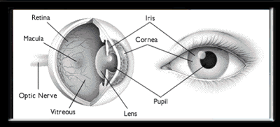
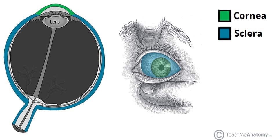
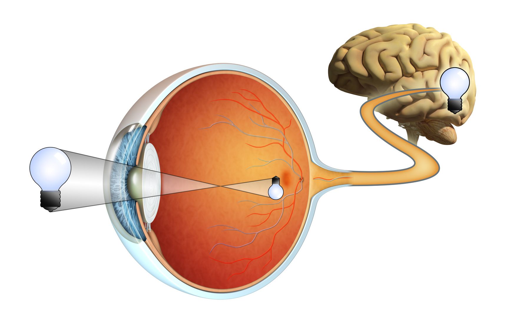
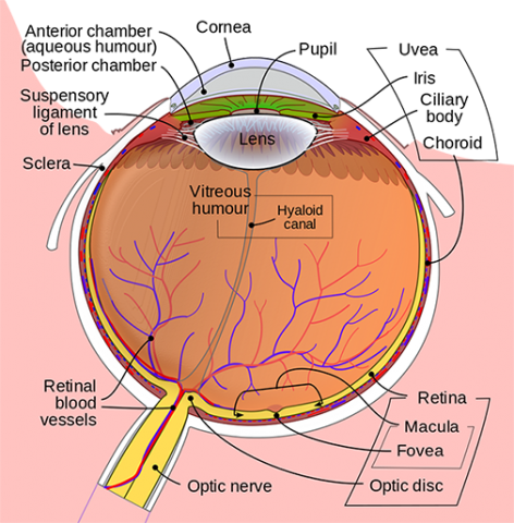
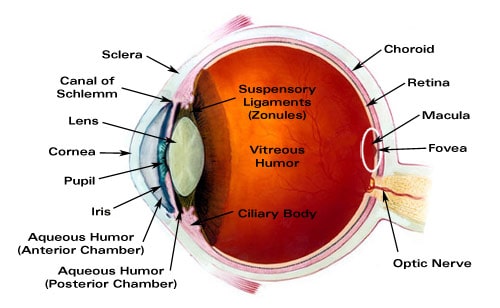
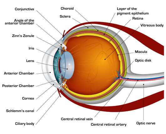
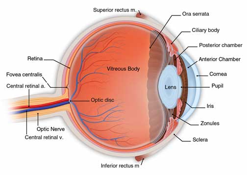
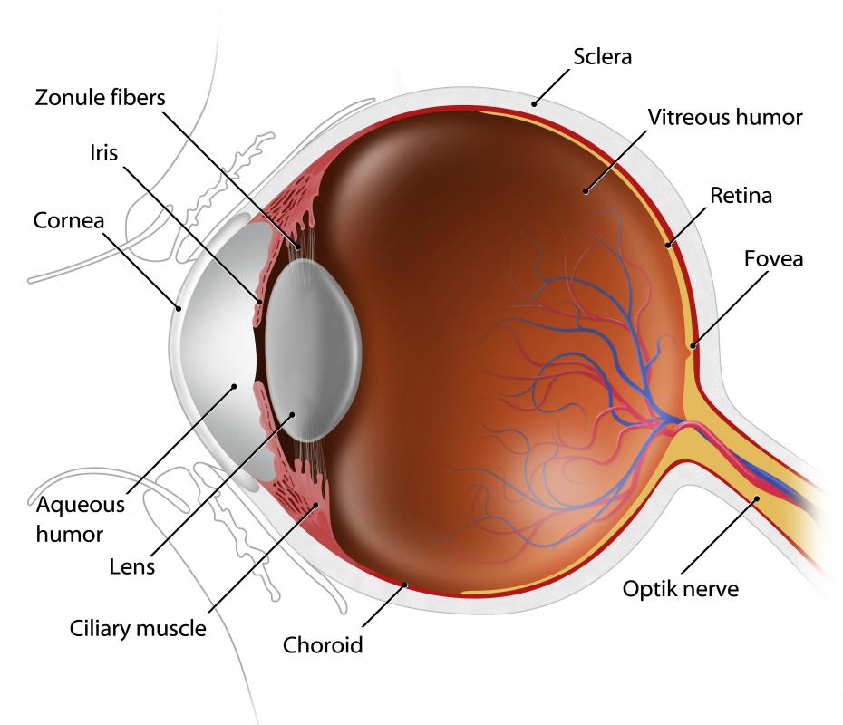
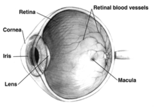
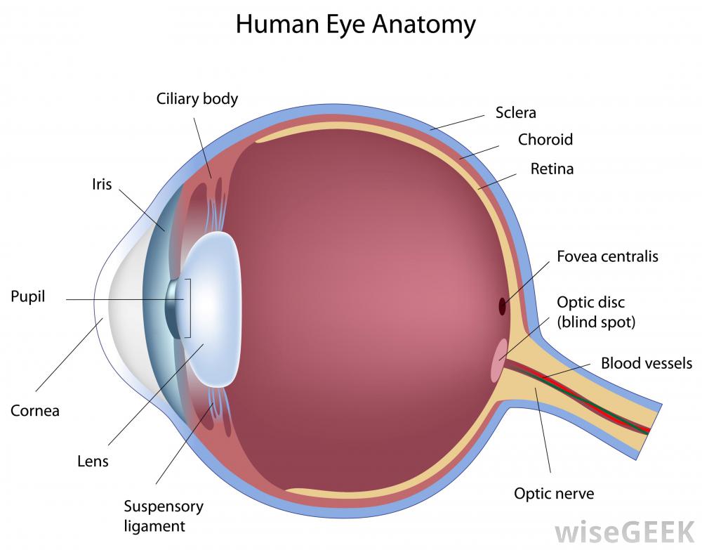
Layers of the eye in order. The outer layer the outer layer contains the sclera the white of the eye and the cornea the clear dome at the front of the eye. These layers lie flat against each other and form the eyeball. So the basic rule is that the top layer will be visible.
And the inner layer of photoreceptors and neurons called the nervous tunic which consists of the retina. It lies in. While dividing the eye into multiple layers can be done in a variety of ways one way to think of layers of the eye is to consider the eyeball as being composed of three main layers.
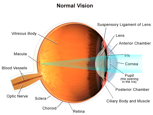
The structure of the mammalian eye has a laminar organization that can be divided into three main layers or tunics whose names reflect their basic functions. The eye is made up of three layers. The fibrous tunic also known as the tunica fibrosa oculi is the outer layer of the eyeball consisting of the cornea and sclera.
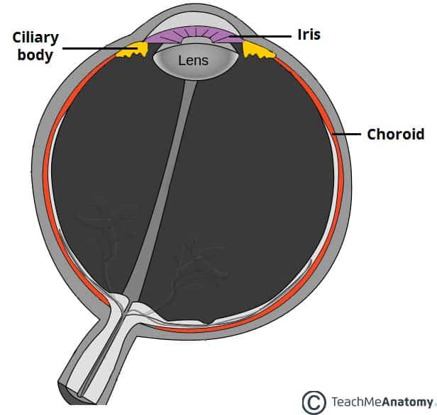
The cornea and sclera. The fibrous tunic the vascular tunic and the nervous tunic. The middle layer responsible for nourishment called the vascular tunic which consists of the iris the choroid and the ciliary body.
The iris choroid and ciliary body. The human eye is a marvel of anatomy providing us with the ability to see the world in all its textures colors and sizes. The outer layer called the fibrous tunic which consists of the sclera and the cornea.
The inner layer of the eye is formed by the retina. Change order of layers. The middle layer is the choroid.
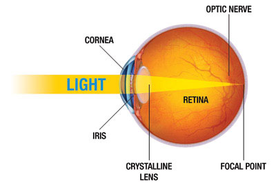
The slight bulge in the sclera at the front of the eye is a clear thin dome shaped tissue called the cornea. The cornea is like a window into the eye. The eye has three main layers.
Start studying layers of upper eyelid. The eye is composed of three layers each of which has one or more very important components. Pigmented outer layer formed by a single layer of cells it is attached to the choroid and supports the choroid in absorbing light preventing scattering of light within the eyeball.
The outer layer of the eyeball is a tough white opaque membrane called the sclera the white of the eye. Its light detecting component the retina is composed of two layers.
 What Does The Eye Look Like Diagram Of The Eye Boozmanhof
The Cellular Structure Of The Retina Infographic Lifemap Discovery
Cow Eye Labeled Diagram Clipart Best
 Vision And The Eye S Anatomy Healthengine Blog
Simple Anatomy Of The Retina By Helga Kolb Webvision
 Anatomy Of The Eye The Ottawa Hospital
The Story Of The Eye The London Project
 How The Human Eye Works Cornea Layers Role Light Rays
 Anatomy Of The Eye The Ottawa Hospital
Https Encrypted Tbn0 Gstatic Com Images Q Tbn 3aand9gcqwr2fcdgdkwkeqcqdwtligrrxpfrz M3sgohmzqjqxus 5n85n Usqp Cau
 The Eyeball Structure Vasculature Teachmeanatomy
 Anatomy And Function Of The Eye
Parts Of The Eye American Academy Of Ophthalmology
 Anatomy Of The Eye Kellogg Eye Center Michigan Medicine
 Major Ocular Structures Laramy K Independent Optical Lab
 Eye Anatomy Detail Picture Image On Medicinenet Com
 The Eyeball Structure Vasculature Teachmeanatomy
 How The Human Eye Works Cornea Layers Role Light Rays
Retina And Vitreous Ophthalmology University Of Kansas Medical
 Vision Optique Layers Of The Tear Film
 Retina Consultants Of Western New York
Parts Of The Eye American Academy Of Ophthalmology
 The Order Of The Three Layers Of Cells In The Reti Toppr Com
Gross Anatomy Of The Eye By Helga Kolb Webvision
 What Are The Different Layers Of Eye Tissue With Pictures
Cornea Research Foundation Of America How The Eye Works

عینک Eyewear Eye Anatomy آناتومی چشم
 Anatomy Of A Normal Human Eye Amdf



Posting Komentar
Posting Komentar