Layers Of The Eye
Retinoschisis refers to the separation of the layers of the retina. The cornea and sclera.
Clearly Com Bay Eyes Laser And Cataract Center Fairhope Al
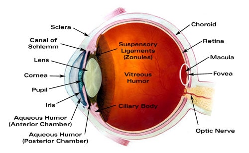
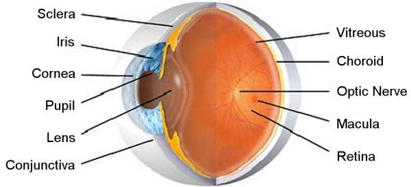
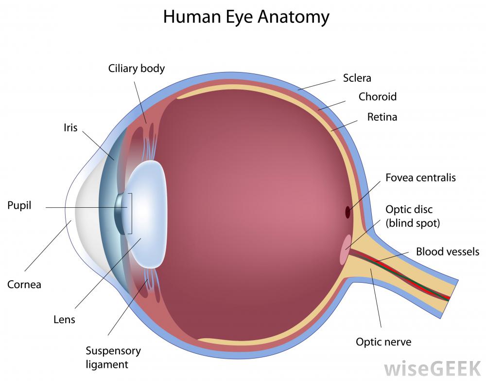
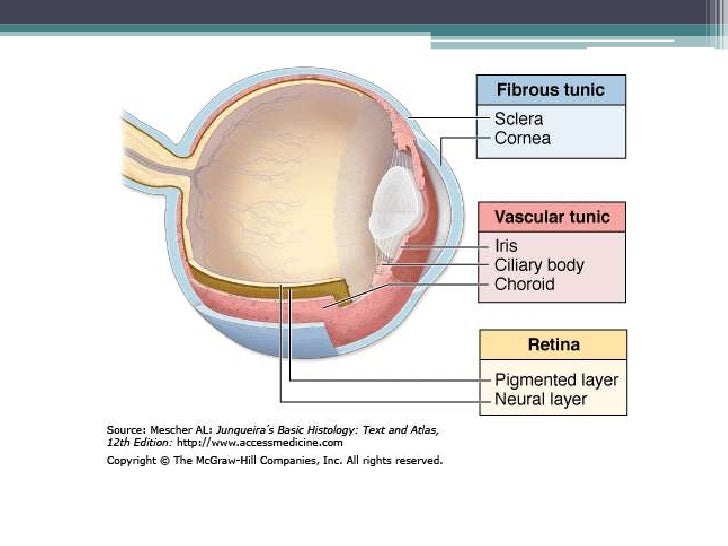
The middle layer responsible for nourishment called the vascular tunic which consists of the iris the choroid and the ciliary body.

Layers of the eye. The fibrous tunic the vascular tunic and the nervous tunic. One eye sees better than the other so your brain favors that eye. The outer layer the outer layer contains the sclera the white of the eye and the cornea the clear dome at the front of the eye.
The structure of the mammalian eye has a laminar organization that can be divided into three main layers or tunics whose names reflect their basic functions. It lies in. Human eye specialized sense organ in humans that is capable of receiving visual images which are relayed to the brain.
There are related clues shown below. The eye is made up of three layers. Eyes are organs of the visual system they provide animals with vision the ability to receive and process visual detail as well as enabling several photo response functions that are independent of vision eyes detect light and convert it into electro chemical impulses in neurons in higher organisms the eye is a complex optical system which collects light from the surrounding environment.
The cornea is like a window into the eye. A problem with the curve of your cornea. The retina is the tissue inside the back of the eye that changes what you see into electrical signals that travel to the brain.
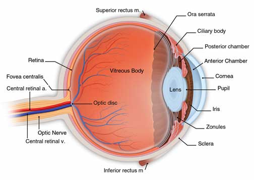
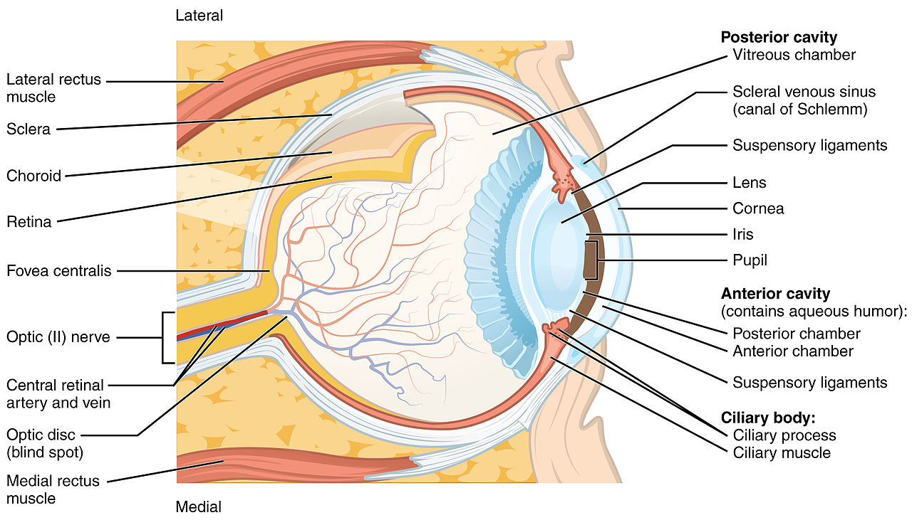
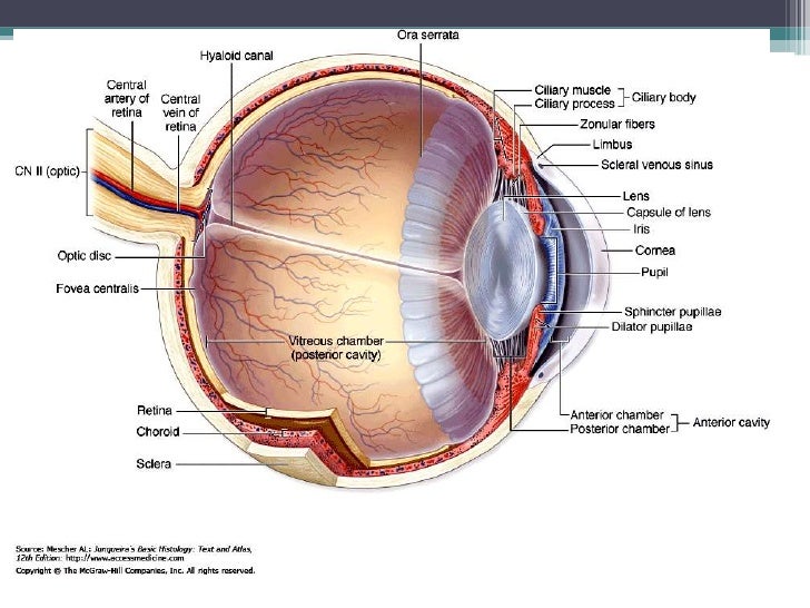
The human eye is a marvel of anatomy providing us with the ability to see the world in all its textures colors and sizes. The iris choroid and ciliary body. The fibrous tunic also known as the tunica fibrosa oculi is the outer layer of the eyeball consisting of the cornea and sclera.
Eye layer is a crossword puzzle clue that we have spotted over 20 times. The eye is composed of three layers each of which has one or more very important components. While dividing the eye into multiple layers can be done in a variety of ways one way to think of layers of the eye is to consider the eyeball as being composed of three main layers.
The outer layer called the fibrous tunic which consists of the sclera and the cornea. The anatomy of the eye includes auxillary structures such as the bony eye socket and extraocular muscles as well as the structures of the eye itself such as the lens and the retina. The weaker eye which may or may not wander is called the lazy eye astigmatism.
And the inner layer of photoreceptors and neurons called the nervous tunic which consists of the retina. Eye layer is a crossword puzzle clue.
 Vision Optique Layers Of The Tear Film
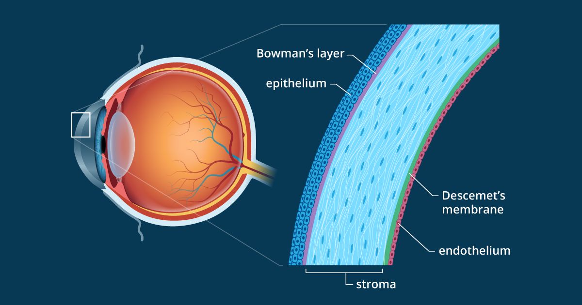 Cornea Definition And Detailed Illustration
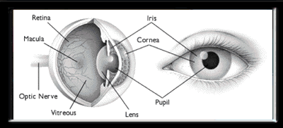 Anatomy Of The Eye The Ottawa Hospital
Eye In Cross Section Anatomy The Eyes Have It
 The Cornea Is Comprised Of 3 Layers Of Tissue
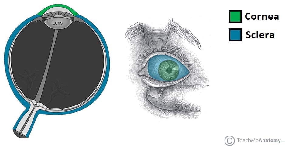 The Eyeball Structure Vasculature Teachmeanatomy
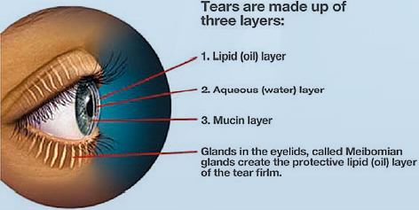 Dry Eyes Sarasota Dry Eye Bradenton Palm Coast Eye Mobile
 Layers Of Eyeball Explained Youtube
 3 Layers Of Eyeball Diagram Quizlet
 Major Ocular Structures Laramy K Independent Optical Lab
How Many Parts Are In A Human Eye Quora
The Anatomy Of The Eye Showing The Three Main Layers Retina
The Anatomy Of The Eye Alison Lawson Centre
 Scientists Discover A New Layer In Human Eye
 Class Eye Structure Online Presentation
 Anatomy Of The Eye The Ottawa Hospital
 Priyamvada Birla Aravind Eye Hospital
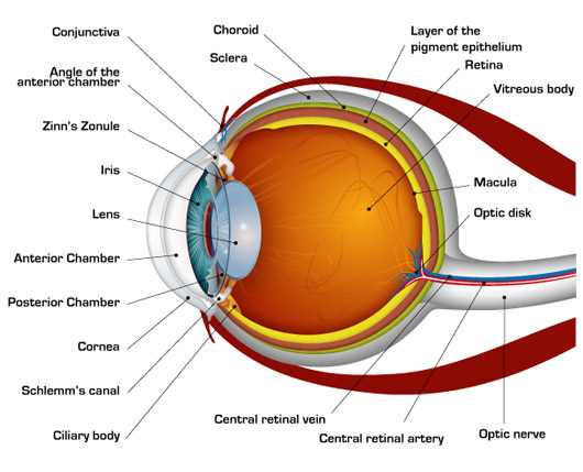 How The Human Eye Works Cornea Layers Role Light Rays
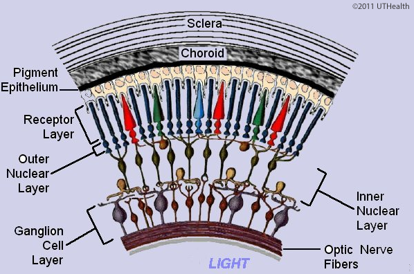 Neuroanatomy Online Lab 7 Visual System Microscopic Anatomy
 Segment And Layer Of Eye B Sc Nursing Youtube
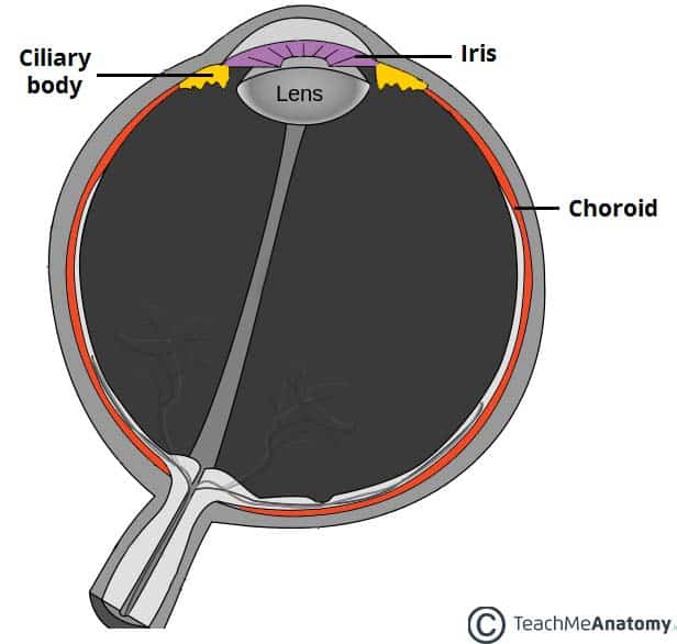 The Eyeball Structure Vasculature Teachmeanatomy
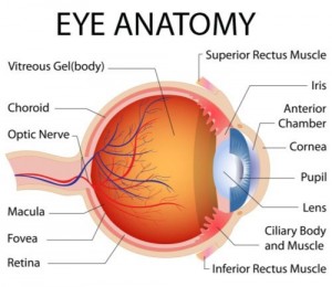 Cornea Center Arizona Eye Specialists Eye Care Phoenix
 File Three Main Layers Of The Eye Png Wikimedia Commons
Https Encrypted Tbn0 Gstatic Com Images Q Tbn 3aand9gcr9fxw98qywsi5kd0skriki0rr5surthojpn3vdptxrenryrir3 Usqp Cau
 Eye Layers Art Print Barewalls Posters Prints Bwc23610275
 Retina Consultants Of Western New York
Human Eye Anatomy Infographic Lifemap Discovery





Posting Komentar
Posting Komentar